DISMANTLING A MYTH:
The Role of Fat and Carbohydrates in our Diet


by Dr. Med. Wolfgang Lutz


ISBN 3-921500-24-9 © 1986 by Selecta-Verlag Dr. Ildar Idris GmbH & Co. KG Planegg V. Muenchen, West Germany

Original title "Leben ohne Brot", translated by Beatrice Idris-Duncan and Joy Wieser. All rights reserved.

No part of this text may be reproduced in any form or by any means electronic or mechanical, including photocopying, recording, or by any information storage and retrieval system, without permission in writing from the publisher.


Service to Danish readers: Danish translation of certain difficult words and medical terms


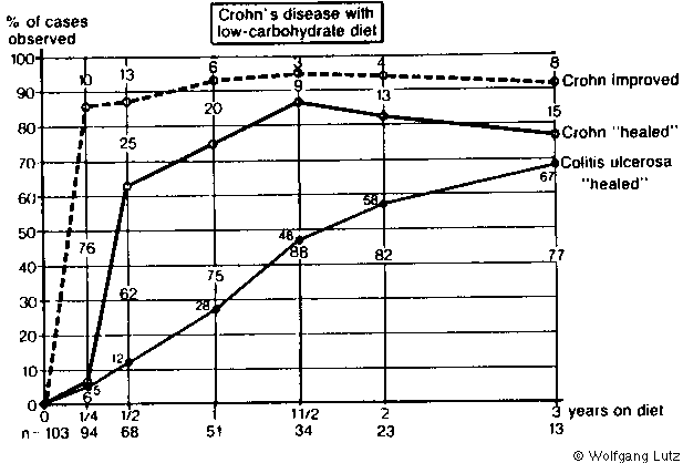
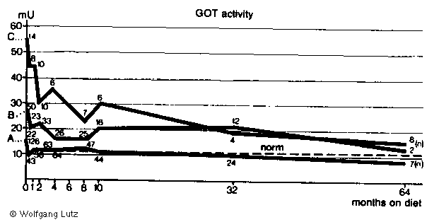
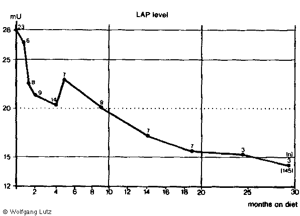
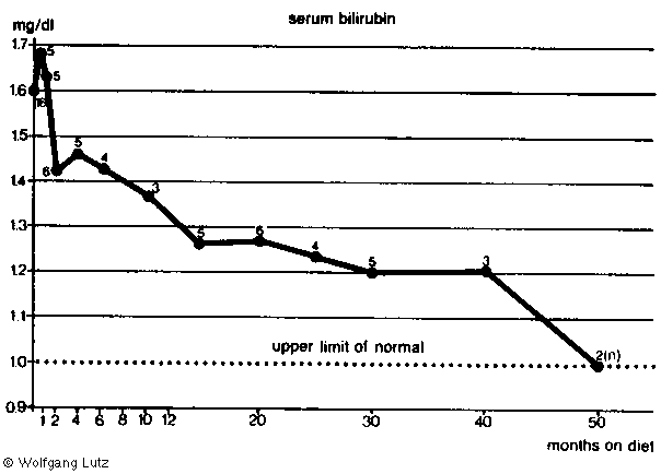
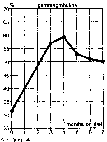
(Excerpt 2: page 125-180)


Chapter VII: GASTRO-INTESTINAL TRACT


To anyone holding the view that carbohydrates are un-natural and harmful components of our diet it is quite obvious that the gastrointestinal tract, being in the front line, is exposed to the most danger. The idea of introduction of a low-carbohydrate diet as a general mode of therapy for disorders of this system is therefore logical, even if contrary to what has hitherto been practiced. Although it has been recommended that refined sugar and white flour be avoided in some gastrointestinal conditions (*1-2), and gliadin-containing types of grain in another (*3-6), carbohydrates as a whole so far have not been considered as undesirable energy sources.


Too Much Acid

Distressing heartburn is often the first symptom to disappear following withdrawal of carbohydrates from the die However severe, and even if made worse by factors like the back-flow of gastric juice into the esophagus in hiatal hernia, the chances of success are good. If patients come back with the complaint that the diet is no longer effective and their heartburn has returned, a closer look usually reveals that some carbohydrates have again crept into the diet. Or a gastrointestinal infection cam be suspected - and treated.

In some way carbohydrates appear to disturb acid regulation, i.e. the normal state of affairs in which acid is produced by the stomach only when it has something to digest. Only a sick stomach produces digestive juices when empty. This so-called 'fasting secretion' is the reason for the auto-digestion seen in gastric ulcers, or at least in ulcers near the pyloric sphincter, the duodenum or at the artificial exit of a resected stomach. Excess gastric acid is responsible for or provides the right conditions for, for development of a gastric ulcer, which is deducible from the fact that a typical gastric ulcer is found only in sites where contact with gastric juice is possible.

If the stomach is resected so that no more acid can be produced a gastric ulcer is permanently cured. In rare cases, however, yet another ulcer develops even if further portions of the stomach have been removed in a second and even third operation. Surprising quantities of acidic gastric juice can be obtained from such patients by means of a stomach tube even in the absence of a test meal or any food whatsoever.

Laboratory tests on such juice reveal that the fasting secretion of acid, just as in patients with a duodenal ulcer, approaches the so-called maximum secretion. By this, we mean the quantity of hydrochloric acid produced by the gastric glands when they are strongly stimulated by food or histamine. Under these circumstances the Zollinger-Ellison syndrome (*7-10), should be suspected, a condition first described many years ago by two Americans, Zollinger and Ellison. Tumors or tumor-like growths are found either in the islet organ itself or in its immediate vicinity, and can be removed surgically.


Gastrointestinal Hormones

In histological preparations of resected material from the above cases, cells were found closely resembling the alpha-cells of a normal islet (pancreas' Islets of Langerhans organ (*11-12). Large quantities of a substance extracted from them proved to be highly effective in causing acid secretion, and was finally identified as 'gastrin' (*13).

Gastrin is the hormone responsible for gastric secretion. Although not the sole agent eliciting acid secretion it is certainly the most important. I do not intend to go into the physiology of gastric secretion in detail since it would take us too far from our main theme. However, it should be mentioned that apart from nervous influences, which for example cause our mouths to water at the sight or smell of food, or even at the sound of the dinner gong, it is gastrin which sets off the flow of gastric juice at meal-times (*14-15). The substance is produced at the base of the mucous glands of the pylorus when the latter is in contact with food. It enters the blood stream and thus arrives at the glands in the stomach's body where hydrochloric acid and protein-splitting enzymes (pepsin, cathepsin) are produced, and where it can directly and indirectly influence the stomach movements, particularly the emptying mechanism.

Thus the stomach is provided with a kind of self-regulating mechanism. Gastrin is formed when food enters, and this leads to secretion of the enzymes and hydrochloric acid necessary for digestion in the stomach. When the stomach contents have been sufficiently acidified the sphincter opens and the acid contents enter the duodenum. Here substances are produced (secretin, cholecystokinin, and enterogastrin for fatty foods) which stimulate the activity of the pancreas and also affect the neutralization of the acid from the stomach, at the same time arresting the efflux of further acidic stomach contents.

The important point to note is that too much gastrin is produced in all cases of hyperacidity, in gastric ulcers and particularly duodenal ulcers, and in extreme quantities in the Zollinger-Ellison syndrome. But this gastrin arises in the pancreas and not in the stomach (*13), (*16-23).

In recent years the so-called Verner-Morrison (*24-29), syndrome has been considered to be a mirror image of the Zollinger-Ellison syndrome. The former is associated with diarrhoea, low potassium levels in the blood, and with a deficiency in free gastric acid. Small tumors contain abnormally-large amounts of secretin (a hormone similar to gastrin but with a different effect) may be found in the pancreas. The two syndromes, however, are not sharply distinguishable from one another as this description would suggest. The symptoms can occur in a variety of combinations, and may be accompanied by disturbances of a diabetic type such as elevated blood sugar or too little sugar (hypos), just as in the case of tumors of the beta-cells.

The patients often complain of headaches and hot flushes in connection with eating, and in fact at such times it is possible to detect abnormally-high amounts of serotonin in the blood and urine. The serotonin is a hormone believed to be formed in the so-called light cells of the islet organ of the pancreas. Some investigators consider them the source of all Islet cells (*30-33).


The Pancreatic Duct Syndrome

In order to understand how the activity of the various segments of the gastrointestinal tract is controlled we have to go back to what we said earlier about neurosecretion in connection with the hypophysis. The nerve cells of the hypothalamus secrete polypeptide substances which reach the hypophysis via small blood vessels and are then either released into the blood when needed, or stimulate the hypophysis itself to produce protein hormones such as the gonadotropins, ACTH, human growth hormone, thyrotropic hormone, etc.

The closer we delve into the problem of neurosecretion the clearer it becomes that it is not confined to the hypothalamus and hypophysis but is in fact a widespread phenomenon. Communication between nerve cells is effected by means of chemical substances known as neurotransmitters. In this way one nerve cell can contact another, its messages can be conveyed either within an organ by means of peptide hormones, or over larger distances by means of the proteohormones which are produced in certain endocrine organs at the behest of peptide hormones. The gastrointestinal tract seems to be especially influenced by peptide hormones formed on the spot by neurosecretion.

The neurosecretory centre is situated in the pancreas in round, glandular structures termed "islets" (of Langerhans), which apparently secrete insulin directly into the blood since there is no sign of effluent ducts. Insulin is an antagonist of glucagon. The large number of different types of cells in the islets would suggest that they form still other, as yet unrecognized, incretions.

In addition to the hormones of the pancreas a larger number of more or less well-known polypeptides with their own regulatory functions is produced in the stomach and intestine. Gastrin is one of them, and although normally produced in the cells of the stomach and reaching its target organ, the gastric mucosa, via the blood stream, it can under certain circumstances also be produced in the small intestine and in the islets of the pancreas. As has already been mentioned, the islets are also the source of secretion.

Since the development of a highly sensitive method for detecting polypeptides, the RIA or Radio Immune Assays, more of this group of substances have been discovered. They are apparently concerned with governing the activity of the gastrointestinal tract and its glands, and include cholecystokinin or pancreokinin, enteroglucagon (an insulin antagonist like pancreatic glucagon), and the gastroinhibitory polypeptide (GIP) from the duodenum and upper small intestine, which is chemically very similar to secretin and glucagon. GIP inhibits the formation of gastric acid and promotes insulin production in carbohydrate digestion, and is thus insulinotropic. It has already been mentioned as representing the probable starting point of diabetes and the reason for hyper-insulinism in carbohydrate eaters. Yet another gastrointestinal hormone is the so-called Vasoactive Intestinal Polypeptide (VIP). It leads to hot flushes and diarrhoea if produced in excessive amounts.

Originally, i.e. in primitive forms of life, the entire gastrointestinal tract was probably able to produce these regulatory polypeptides. In the course of further developments, however, the various parts took on a specialized function.

Esophagus, stomach, duodenum, small and large intestine, differ not only in structure but also in function, so as to serve the body in the best possible way. In the course of this transition glandular organs have fused, in order to produce the liver and pancreas, whose only present connection with the intestine is via the ducts through which their secretions enter its lumen. As has already been mentioned, the neurosecretory portion of the pancreas is concentrated in the islets.

Neurosecretion is nevertheless not confined to the pancreas and its islet organ, but still plays a role over the whole of the gastrointestinal tract. Even though insulin and glucagon are apparently produced exclusively in the islets the other gastrointestinal hormones can be produced elsewhere too, like gastrin, at least under pathological conditions.

In the embryo the primitive pancreatic duct from which the pancreas develops appears to harbour the primitive form of every kind of cell producing gastrointestinal hormones. With this in mind I suggested the term "pancreatic duct syndrome" for all disorders triggered by gastrointestinal peptides.

The cells producing gastrointestinal hormones can be thought of as belonging to a clan dating back to primitive times. Some members of the clan have built larger houses (the islets of the pancreas) whereas others still live in the open or in very primitive shelters.

Nonetheless the clan sticks together through thick and thin, and their sense of loyalty is such that if one of the members is injured, an outcry will be heard from the rest. Emanation or irradiation is a familiar idea to members of the medical profession.

Apparently a strong stimulus to one gastrointestinal polypeptide or to its site of production involves to greater or lesser extent all of the others. Not only is insulin or glucagon produced, but gastrin, secretin, serotonin, GIP, VIP, as well, and probably others as yet unrecognized.

That the disturbances already discussed are due to the carbohydrates in the diet, and that they disappear if carbohydrates are restricted, is probably due to the fact that the two chief hormones of the islet organ, insulin and glucagon, primarily regulate carbohydrate metabolism. They respond to too much or too little blood sugar, and the presence of carbohydrates in the intestine. And least one of the gastrointestinal polypeptides (GIP, VIP) has an insulinotropic action.

It was previously assumed that sugar injected direct into the blood necessarily has a greater effect than the same quantity administered orally. Surprisingly, the opposite holds true and sugar in fact is much more effective if it enters the body via the intestines, i.e. insulin production is stepped up much more rapidly and is more persistent than if the sugar is given intravenously.

The reason for this is I think that when sugar enters the intestine insulinotropic substances are produced the mucosa and as their name suggests they stimulate the pancreas to produce insulin. They are the outposts which serve to warn the headquarters that sugar is on its way, and that insulin has to be produced for its breakdown.

It might be that these outposts represent the seat of our civilIzation diseases (see also chapter XIII). Our primitive ancestors learned to live without carbohydrate over the course of millions of years and now our body, via the above-mentioned outposts, reacts "allergically" to the kind of food that was introduced with grain cultivation, and even more to the large quantities of sugar that we have consumed since the development of sugar-beet cultivation.

The 'clan' reacts as to an enemy against which it is in fact no longer armed. It throws in all the resources at its command even if the weapons involved are not suited to the purpose. The body produces not only insulin and glucagon but secretin, pancreozymin, vasoactive intestinal polypeptides, and above all gastrin as well. Like a beleaguered garrison it finally resorts to producing ammunition in workshops originally intended for quite different purposes. Similarly, as a result of prolonged excessive intake of carbohydrates, gastrin is finally produced by the pancreas as well as by the stomach.

That this somehow describes the situation is clearly borne out by the observation that all symptoms connected with the pancreatic-duct syndrome disappear upon withdrawal of carbohydrates from the diet. Although I have not yet had the opportunity to put my idea into practice I am convinced that the Zollinger-Ellison syndrome would, if not the result of a malignant tumor, eventually respond to a low-carbohydrate diet if continued long enough, just as does Morbus Cushing.

There is little doubt that in addition to GIP or VIP the typical fluctuations in the blood-sugar level and the attendant counter-regulation so typical or carbohydrate consumers play a special role. This was discussed in the chapter on diabetes and hypoglycemia. It is striking that in cases of hyperacidity and duodenal ulcer the maximum acid production in connection with eating is the same as in normal subjects, whereas the so-called basal secretion in the fasting state is higher than normal. And it is in this fasting state that the tendency to hypoglycemia becomes manifest, with its resultant need for increased glucagon (and gastrin) production.

HIstological similarities between the gastrin-producing cells in tumours from Zollinger-Ellison patients and the glucagon-producing alpha-cells of the islet organ support this theory. Viewed in the light of such observations it would appear that gastric surgery is being performed in the wrong place. The removal of the lower part of the stomach (antrum) deprives the organism of a gastrin production geared to the requirements of the digestive tract, whilst leaving the cells that are active between meals to produce the fasting secretion and ulcers.

I am convinced that we would spare many of our patients the need of undergoing surgery if we were to put them on a low-carbohydrate diet. An operation is, after all, only a form of mutilation leaving the root of the trouble untouched.


Gastritis, Enteritis, Ulcers

The above ideas have been fully confirmed by my experience on the effect of a low-carbohydrate diet in patients with hyperacidity and duodenal ulcers. Both gastritis and ulcers heal if carbohydrates are restricted. A so-called callous gastric ulcer, however, should not be treated immediately by simple restriction of carbohydrates. These ulcers are stress-induced and a transition to a new diet entails further stress. Therefore one has to give small amounts of cortisol first until the ulcer has healed and then slowly introduce a low-carbohydrate diet.

In my experience the only permanent relief for an ulcer lies in consistent adherence to a low-carbohydrate diet. Lack of discipline in this respect usually leads to the operating table.

The frequent mention of ulcers in connection with gastro-intestinal disorders does not necessarily mean the condition is as commonly encountered in a physician's practice as in a hospital, where there is simply an accumulation of cases that cannot be treated at home. Even the duodenal ulcer is a relatively rare condition, at any rate less common than gastritis, enteritis and colitis.

It was originally believed that mucosal irritations could be detected by means of an X-ray. More recently, however, biopsy methods have been developed in which small pieces of mucosa are removed from the stomach or intestine. Their histological examination has provided us with more detailed knowledge as well as some surprises.

We know that the story begins with excessive irritation of the mucosa and increased production of gastric acid and digestive enzymes.

At this stage the mucosa presents a fairly normal aspect under the microscope. X-ray pictures show the so-called fasting secretion, i.e. even an 'empty' stomach, is full of juice. The mucosal folds are either normal or swollen, indicating that adequate substance is still available. That this type of gastritis represents the initial stages of the disease and is not another variation of it is demonstrated by the observation that it is the common form in children and young subjects where the disorder is not long-standing. X-ray pictures of children with gastritis almost invariably reveal normal or enlarged mucosal folds and a copious fasting secretion.

The longer the process has time to develop the less fasting secretion is seen. The sphincter is open and the stomach is seen to empty its contents prematurely into the small intestine. In the end, what was initially an excess of gastric acid and pepsin is now too little and in some cases NO gastric acid can be demonstrated at all. An experienced eye recognizes this in an X-ray picture, but it becomes very obvious in pieces of mucosa examined under the microscope that atrophy of the mucosal layers has set in and large numbers of mucosal cells have perished. Heartburn and other symptoms of hyperacidity should not be allowed to mislead one. Even with such a thin mucosa the stomach can still produce acid in quantities sufficient to cause a severe burning sensation in the lower oesophagus, whereas the normal stomach moves its contents in a downward direction (except in vomiting), a diseased stomach can in some cases regurgitate its contents into the oesophagus.

The mucosal atrophy, whether due to chronic damage, or --- as mentioned earlier --- to excessive secretin (GIP, VIP) production which might to be responsible for the deficiency of acid, leads to a series of consequences for the digestive apparatus. Raw connective tissue, in the absence of adequate amounts of acid or pepsin, is insufficiently broken down and even the juices from the liver and pancreas in the small intestine are unable to complete the process. Such patients often suffer from diarrhoea if they eat raw or smoked meat. This is no great misfortune and the patient can easily avoid such foods if made aware of the necessity.

The deficiency of acid, however, results in malfunction of the emptying mechanism of the stomach. Normally the gastric sphincter shuts as soon as the acid content in the duodenum rises. In this manner the passage of further acid contents is delayed until the acid has been neutralized by the alkaline juices of the duodenum (bile and pancreatic juice). If the stomach produces too little acid the point of neutralization is reached sooner, with the result that it empties too rapidly. The stomach thus loses its function as a depot and the rest of the digestive tract is overburdened by receiving insufficiently-digested food.

But breakdown of food is not the only function of the stomach. For example, it produces a substance, the Intrinsic Factor, which by combining with vitamin B-12 (Cobalamine) in the food, and perhaps with other so far unknown substances, renders it absorbable. If the mucosa is atrophic and unable to form intrinsic factor, the vitamin is not absorbed but is lost in the stool. Since vitamin B12 is essential for growth of the entire organism as well as for maturation of the red blood corpuscles it is understandable that patients of this type suffer from a variety of disorders including pernicious anaemia.

Although no-one believes today that gastritis and enteritis are expressions of bacterial or viral irritations, we still have no exact idea as the cause of these common complaints. In my opinion we are dealing with nutritional diseases provoked by the high carbohydrate content of the diet of civilized man. Carbohydrates elicit production of gastrin, secretin and so on, and in the initial stages the mucosa is stimulated to produce excessive quantities of digestive enzymes, and this at the wrong time, until finally it succumbs. The process is enhanced by the tendency to tissue deterioration seen in carbohydrate-eaters. Every organ has to make a contribution to the toll of protein that is constantly demanded of carbohydrate-eaters by the mechanism of the adrenocortical hormone. The process is the same as that already discussed in connection with the striae.

I have described these processes in detail because their understanding is of vital importance in treating such patients. The more recent the process the earlier the patient comes for treatment, the more fasting secretion, enlarged mucosal folds and other signs of mucosal swelling and hyperacidity, the sooner can the patient expect to be relieved of his complaint by means of a low-carbohydrate diet. The better-established the process, the thinner the mucosa and the more rapid the stomach emptying the longer it takes to effect a cure, and in some cases the less complete will this cure be. But an improvement can be expected in any case.

Chronic inflammation of the stomach (gastritis), the duodenum and small intestine (enteritis), and large intestine (colitis), often occur together, although in most cases the symptoms of one or another of the organs or hormones of the pancreatic duct system usually dominate the picture. The characteristic symptoms of gastritis are a feeling of sickness, nausea and vomiting in the fasting state, usually tine morning and following breakfast, choking, a lump in the throat and a burning sensation the stomach. Fasting pains that are usually improved by food to return as after-pains a couple of hours later suggest disease of the gastric pylorus or duodenum. Enteritis is usually accompanied by a full feeling after meals, rumblings. gas, inability to digest fats and colicky stomach pains.


Fat is not the Culprit

Because fat in the diet evokes unpleasant symptoms in many patients, the opinion has arisen that a gastric diet has to be low in fat. But a low-fat diet never brings about a cure. In fact the diet that I recommend for patients can later include large quantities of fat. The patients feel distinctly better even within a few days and usually are completely well again within a couple weeks. But if they attempt to eat carbohydrates they quickly notice that these were responsible for the discomfort and not the fat or other foods.

In very severe cases of enteritis the small intestinal loops are knotted and swollen, and the patients have a pot-belly similar to that seen in primitive peoples whose diet consists mainly of carbohydrates. The children often already suffer from Kwashiorkor, a disease characterized by a state of malnutrition, swollen belly and oedema, and resulting from their one-sided, low-protein diet. Even in civilized countries we find equivalents to this disease in which, for unknown reasons, dietary fat cannot be utilized but is eliminated in the stool.

A typical disorder of this kind is celiac disease in infants which results from oversensitivity to the protein gliadin (*3-5), a substance present in wheat, rye, barley and oats. Since corn, rice and potatoes contain none of it, it is quite a simple matter to rear the patients on a gliadin-free diet and at the same time ensure their normal development.

The absence of the necessary enzyme for splitting gliadin from the intestinal villi suggests that the gliadin sensitivity is of genetic or immunological origin. A gliadin-sensitive type of enteritis is also encountered in adults in the condition known as endemic (as opposed to tropical) sprue (*5). In this disorder, as in other forms of chronic enteritis, not only can antibodies to gliadin or its individual fractions (e.g. fraction III) be demonstrated, but also antibodies to hens' eggs, milk, and even to rabbit protein (*34-36).

It can hardly be assumed that the defect is of genetic origin in these patients but rather that the damaged mucosa permits the passage of antigens which elicit the production of antibodies in the blood. This would mean that the mucosal damage represents the first step in the disease and can thus be regarded as the cause and not the effect of auto-immunization.


Itching (Pruritus)

I have another reason for believing that this is indeed the situation. A large number of patients visit the doctor on account of itching, which they report as occurring after wetting of the skin (bathing or sweating), and also after eating certain food. If these patients are put onto a low-carbohydrate diet the itching usually disappears after a couple of months. My explanation is that the diet gives the intestine a chance to heal, the mucosa recovers and no longer lets through (or completely breaks down) the food constituents that could previously enter the blood and elicit an allergy.

Since I have been so much concerned with the clinic and radiological signs of enteritis I have come to realize just how many apparently clinically healthy patients suffer in this way. Only when closely questioned do they admit their discomfort, and for this reason the argument that such antibodies are found in 'healthy' subjects and has nothing to do with enteritis does not hold water.

It is small wonder that the gliadin-free diet automatically prescribed for adult enteritis is not particularly successful.

When the condition due to low-carbohydrate has improved to the point where the patients can safely begin to add again some carbohydrate to their diet it is bread and other flour-containing foods that are least-well tolerated. I believe that this is not because gliadin is responsible for the enteritis but because it has a greater allergic effect than other unsplit proteins if it gets into the blood. Therefore bread Is forbidden to such patients. They may replace the carbohydrate with fruits, vegetables, potatoes and pulses.

The fact that carbohydrates have something to do with gastro-intestinal ailments was already known to Prof. H. Lampert, who recommended restricted carbohydrate intake for the so-called fermentation dyspepsia (when stools are acidic and smell peculiarly acidulous), and for abdominal flatulence (*36). Had he continued on this path, he surely would have observed that not only "fermentation dyspepsia" but also "putrescent dyspepsia" responds to carbohydrate restriction.

For the latter disorder he recommended other diets.


Colon Disorders

Constipation

On the whole, constipation is the most common illness of the colon, but it is mostly restricted to the female gender since the female hollow organs, which include the uterus, tend to retain their contents in order to preserve the fetus, instead of expelling them. Therefore women are also more susceptible to enervation and infections of the urinary tract, but less susceptible to ureteric colics.

Without laxatives the stool of chronically constipated patients is broken down by putrefaction bacteria. The gases produced during this process can he taken up by the blood and detoxified in the liver. Or the tormented gut finally rids itself of the stools by the so-called paradox diarrhoea.

Billions of dollars are realized by producers of laxatives but the ailment is treated only very superficially. Experience shows that the effect of laxatives diminishes with time so that the dosage has to be increased or the preparation has to be changed. In the long run they are most likely health-detrimental since they cause an evacuation of the bowel by irritating it, and because they have been demonstrated to disturb the mineral metabolism (loss of potassium). This constant irritation of the mucosa finally leads to the most predominant complaint among chronically constipated patient: they feel constipated although their colon is totally empty and laxatives would not be necessary.

When these patients come to the physician, he justly recommends the discontinuation of the medication. But without laxatives the patients have no stool. If they are put on a low-carbohydrate diet then they definitely have no evacuation. Now the last motivating force for the bowel abolished because the fermentation process stops and the entire gastro-intestinal tract calms down. Even the female patients who are not normally constipated react to a low-carbohydrate diet with an indolent bowel. That is why I have not initially treated constipation with this diet. However, over the years I solved this problem by instructing my patients to continue the laxatives and by prescribing them instead daily cleansing enema with one and a half litres of warm water without additives in order to bridge the period of initial change for the worse.

Under the continued diet, sooner or later the stool will normalize. In children it can take one or two days, in young adults one or two weeks, and in older persons a few months, but undoubtedly it will be successful sooner or later.


Diverticulosis

The wall of the colon consists of several muscle 1 which cross each other and, like a lattice, leave open spaces through which blood vessels and nerves pass. When these muscle layers weaken, the spaces enlarge to the point that mucosa can protrude through the meshwork. These finger-like mucosa protrusions get under the peritoneum, which covers most of the colon and becomes deformed into button shapes. These "buttons" are covered by peritoneum on the outside and are therefore fixed in place so that they can no longer retract. On the inside they contain stool or other bowel contents.

The number of these so-called diverticula can vary greatly. There are patients with one or two diverticula and others with 20 to 50. They generally do not cause typical symptoms. Only when they become inflamed does the resulting peritonitis create problems; at times it necessitates immediate surgery. The diverticula are most-commonly localized in an area of the large bowel called the pelvic colon, an S-shaped loop between the rectum and descending colon on the left side. Here an inflammation creates symptoms like an like an appendicitis would be on the right.

Usually a diet rich in fibers is recommended for the treatment of diverticulosis because years ago scientists observed that African Negroes who eat fiber-rich diets have no diverticulosis and also get few other colon diseases (cancer).

I, however, do not believe in this fiber hypothesis which has become very popular over the last few years. Man is a hunter and gatherer and not a herbivore. although his original diet may have contained somewhat more-undigestable food residues. Therefore, I also treat diverticulosis with a low-carbohydrate diet very successfully. Here the prevention of an initial constipation through the regular application of cleansing enemas as described in the previous chapter on constipation is particularly important.

After a few months the patients will have normal stools and no further complaints relating to their diverticulosis. The existing diverticula will not disappear but no new ones will form. Most of all, the inflammation, which very often exists in the gastro-intestinal canal of consumers of normal diets, and which spreads to the diverticula, will heal. Finally, the bowel musculature will strengthen and the original cause of the diverticula, namely the muscular weakness with the enlarged spaces between the bundles, disappears.


Diarrhoea

I am not talking about the short periods of liquid stool which occur several times a year due to infections with various bacteria or viruses. Particularly viruses multiply in the gut, enter the blood stream, and finally lead to a general illness.

A frequent traveller knows that "Montezuma's Revenge" is not restricted to Mexico. On long trips one enters regions with unfamiliar germs to which one is not yet resistant and promptly becomes ill.

Here I mean the chronic diarrhoea, which is much more common than is generally assumed. One just has to visit larger toilet facilities in a restaurant or train station to hear the familiar noise in one of the neighboring stalls. However, this illness is evaluated differently by various people. Some have had diarrhoea for years and have become so used to it that they do not perceive their condition as unusual anymore.

I remember one patient, the foreman of a Mercedes-Re service station, who came to me years ago and told me that he has had diarrhoea for over 15 years now and that he had consulted at least ten physicians without any success. He did not dare to leave his house anymore; when he had to out anyway, he would immediately look for a rest stop since at times he could hold his stool not even for one minute.

I prescribed him a low-carbohydrate diet and told him to see me again in a fortnight, because I was sure that the problem would have resolved by then.

This was indeed the case. He came, thanked me and told me that of the many doctors he consulted previously, not one knew how to help him. He did not quite understand that, because if there is mechanical trouble with one the Mercedes-Benz models, all service stations in the whole world would be informed and instructed how to repair the defect. Whether this was not so in medicine? No, I had to admit: we have the so-called scholarly medicine with certain dogmatic views, and other views are not tolerated. The many "outsiders" among the medical scientists will surely agree with me wholeheartedly.

When chronic constipation is to be understood as disturbed motility of the bowel due to an insufficiently-developed colon musculature, which is unable to move its contents ahead (which Is hardly the heart of the problem), then chronic diarrhoea surely cannot be considered solely as motility disturbance. Rather, the bowel evacuates because it does not want to contain tts contents; it is not able to absorb water and to produce stool of a normal consistency; therefore it releases the contents of the small intestine in a more or less unaltered form.

In my experience, acute diarrhoea is followed by a lengthy period of liquid or soft stool, although through therapy and the elapsed time interval the Infectious organism has been killed off and the Infection has been overcome. Furthermore, this type of diarrhoea can be halted by giving gold or cortisone for several days, i.e. by subduing the immune reaction; therefore we are dealing here with a so-called "auto-aggressive disease".

For reasons which are not yet known in detail, the body starts to mount an immune reaction not only against the infectious organisms but also against the affected organ. I got the Impression that initially this mechanism does not necessarily have to be considered morbid, that the attack against the affected organ is part of the immune reaction itself. The body tries not only to fight the infectious organism, but also to destroy its basis for existence, the affected organ, by weakening it with its defensive forces. One can hardly understand the multitude of existing auto-immune diseases in any other way. These affect not only the gastro-intestinal tract but also the nervous system (multiple sclerosis, see page 239) and, most of all, the heart. The myocardial inflammations, which frequently occur even among young people, can most easily be explained in this way.

When diarrhoea after an acute gastrointestinal inflammation does not heal and persists for years, one not only has to restrict carbohydrate intake, but also to slow down the immune reaction. This can be done with cortisone or gold preparations. The former is less harmful and therefore should be tried first. I prescribe 10 mg Prednisolon or 8 mg Triamcinolon (early in the morning for eight to ten days) and repeat the same treatment in case of a relapse.

Frequently an immunosuppressive therapy is not even necessary. After one or two weeks the stools normalize although often accompanied by abdominal pain. One has to know this in order not to break off the diet, believing it to be inappropriate. Apparently the large bowel, which has not actively worked for months or years as it has discharged the contents coming from the small intestine in a more or less unaltered form, rebels against having to work again.

Sometimes even a period of constipation follows, which should be treated with cleansing enemas, since laxatives should be avoided at all cost. Ultimately, chronic diarrhoea will always heal under the above-mentioned circumstances and with a low-carbohydrate diet.


Crohn's Disease

Why chronic diarrhoea, as described above, develops into a Morbus Crohn (English: Crohn's Disease), named after the American physician Dr. Crohn (*37) is not clear. Apparently there are connections between the two, because in my experience, a "Crohn's" always has a pro-dromal phase in which the patient has diarrhoea over some period. It is, so to speak, nothing else but the extrapolation of the chronic enterocolitis. Here the illness is not limited to the mucosa alone but Involves all levels of the bowel, the mesentery, and the lymph nodes; the gall bladder, the duodenum, and the stomach can be affected as well. A form of InflammatIon, called granulomatous, is characteristic. Infiltrations around the bowel in the peritoneal cavity can result, but they also can melt down, form abscesses, and leave fistulas, through which the bowel contents can enter the bladder, the vagina, or, most frequently, exit next to the rectum or through the abdominal wall. Often they can only be removed surgically and often without ideal results.

Crohn's disease becomes more common from year to year. When I started to treat my colon-patients with a low-carbohydrate diet 25 years ago, there was no Crohn's patient among them. Lately they have become almost as frequent as patients with ulcerative colitis.

Morbus Crohn differs from ulcerative colitis in that there often is no intestinal bleeding, and in its different localization. Crohn's rarely occurs in the rectum; its frequency Increases upward; often it affects only the lower small intestine. Ulcerative colitis, on the other hand, prefers the rectum; its frequency decreases continually toward the small intestine; at least sporadic bleeding occurs and no fistulas form. There are intermediate forms between ulcerative colitis and Crohn's disease: patients with intestinal bleeding and fistulas, those with involvement of the large and small Intestine.

The distinction between Crohn's and ulcerative colitis is of significance. Crohn's disease, in contrast to ulcerative colitis, heals with great probability within six months to one year and usually without complications. There are individual patients who have difficulties changing from the high-carbohydrate standard diet to the low-carbohydrate diet; they worsen acutely, have diarrhoea, fever and so on. However, this rarely lasts longer than a few weeks. Potential remnants (fistulas, etc.) must be removed surgically if they do not heal spontaneously; also narrowings in the bowel may require surgical intervention. However, if possible, one should wait until the underlying illness has resolved, until iron level, erythrocyte sedimentation and stools have normalized.


Fig. 16:
103 patients suffering from Crohn's disease were treated by a low-carbohydrate diet. After a quarter of year most patients (85 percent) showed remarkable improvement in their health. After half a year, more than 60 percent were asymptomatic, after one year more than 70 percent and after one and a half year about 85 percent. This is in contrast with ulcerose colitis, which Is shown in the lowest line, improvement of which runs much slowlier on the same diet and often is interrupted by relapses.

Crohn's disease generally thought of being incurable can so be shown to be very well accessable to dietary measures.


In March 1985 I published 103 Crohn's patients (Fig. 16) which I had treated up to then (*74-75), and with this information I hope to be able to convince my colleagues to first try to treat these unfortunate patients with diet instead of cortisone and scalpel. Perhaps then also a breakthrough regarding other diseases can be reached. In recent years German and English authors (*38-39) have found that Crohn's patients experience a certain improvement after relinquishment of sugar and so-called refined carbohydrates. This corresponds to today's widespread conception that only the refined carbohydrates are harmful, i.e. sugar and white flour.

I do not believe this. To the contrary, I am convinced and have seen again and again that bread and particularly whole meal bread is not tolerated well by patients with gastro-intestinal ailments. Starch turns into sugar in the bowel anyhow, perhaps somewhat slower than if one eats sugar and only dextrose, but in principle the result is the same. At least this recommendation points in the right direction: restriction of carbohydrates.

Often a gluten-free diet is tried, particularly for chronic diarrhoea and Crohn's disease, because the alcohol-soluble fraction of the grain protein causes the so-called Celiac Disease in children, which is accompanied by pulpy, flat-cake-shaped stools, and impaired development. But I have seen many times that grain protein is not the cause of this colon disease. Again and again patients were on a gluten-free diet for months or years, unsuccessfully, but responded to carbohydrate restriction despite of gluten.

Recently the consumption of margarine and other artificial fats as cause for Crohn's disease has been discussed, based on the fact that in France and in the WalIonic part of Belgium, where less artificial fat and more butter is consumed, Crohn's disease occurs less frequently (*40-41).

Indeed, the increase in Crohn's disease in the German-speaking countries in the last years correlates with an increase in margarine consumption. One speculates that the artificial fats are not broken down and absorbed properly, so that their metabolites enter the lymphatic vessels and cause the described granulomatous inflammations. This could be quite true. Artificial fats are, considering human evolution, definitely less natural than carbohydrates, but I have among my patients cases of Crohn's disease who have only eaten natural fats and never artificial fats, but I have none who has eaten no carbohydrates. Yet I can imagine that the consumption of artificial fats supports the tendency to develop the disease, when carbohydrates are consumed in large quantities.


Ulcerative Colitis

Ulcerative colitis is located at the lower end of the continuum "chronic diarrhoea ---> Crohn's Disease". It may begin one day quite harmlessly with the appearance of blood in the stool. The physician diagnoses hemorrhoids which cannot be found with the rectoscope. Instead he sees a reddened mucosa which starts bleeding at the slightest touch or when the overlying purulent layer is removed. In mild cases this process is restricted to the rectum; the more severe the disease the further it extends upward. In contrast to Crohn's Disease the small intestine, the gallbladder, the duodenum, and the stomach are never involved. There are no infiltrates, no perforations, and no flstulas. Only the mucosa and the underlying muscle layer are affected. However, in time both are destroyed to an extent that the whole colon contracts into a short cylinder, which is completely motionless and has lost most of its function.

At least these patients still have their natural anus and not an artificial abdominal one. All this is bad enough but the patients would and actually do put up with their bloody stools, abdominal pain, and diarrhoea, if the disease would not become life-threatening by turning into the so-called toxic megacolon or by degenerating malignantly. A toxic megacolon can develop at any time as long as the ulcerative colitis is active. Only three patients of a total of 285 that I have treated up to the end of 1984 have died. One was a relatively young man who had recovered quite well under a low-carbohydrate diet and had gained 20 kg of weight. Suddenly he became acutely ill with high fever and abdominal pain. He was admitted to a clinic but was operated on too late because he had fiercely refused this operation. Another one underwent surgery without my approval; the third, a very old lady, died two months after operation of thrombo-embolism.


Surgical Therapy

Of all patients with ulcerative colitis 20 to 30% sooner or later have to be operated on if they do not eat a low-carbohydrate diet. The constant loss of blood and iron and the continuously-deteriorating general health necessitate the removal of the colon and the creation of an artificial anus (*42). Only two of my patients had to have their colon removed. Of course I cannot be certain that one or the other who was lost to follow up (who did not answer my repeated inquiries) did not have to have his colon removed after all. Certainly there is a limit even to what a low-carbohydrate diet can do, a stage of the disease so advanced that the transition to a new diet is not tolerated or effective anymore. Maybe the patients gave up after months or years of dieting, or other reasons necessitated surgical intervention.

With the surgical removal of the entire colon the disease is finally overcome. Not only the colitis is resolved because the diseased organ is gone, but with it all other symptoms of a chronic colitis which can occur in the eyes, skin, and the joints. They manifest due to an outward radiation of the immune reaction from the colon to other organs.

There is still one other possibility before the operation, and that is gold therapy. Gold is a well-known anti-rheumatic drug which was used extensively to treat joint inflammations before the newer non-steroidal anti-inflammatory drugs and cortisone preparations became available. Gold is directly taken up by the immune-competent cells, the white blood cells and their progeny; it inhibits their activity and thereby improves immune diseases. The leukocytes are not able to attack the colon's mucosa anymore.

Gold therapy should only be applied to severe cases of ulcerative colitis because sometimes it causes side-effects like week-long fever or urticarial skin rashes. It can also alter the hormonal status. I have observed that the gonadotropin secretion decreases (see page 94), which can compromise sexuality. In women this is not so bad, sometimes even advantageous, because menstrual breast pain disappears. However, in men sexual potency often declines. Of course, in severe cases of ulcerative colitis one will have to ignore these side- effects, since usually the question is whether the colon can be preserved or not. The side effects usually disappear with time.


Low-Carbohydrate Nutrition

I can only report positive about my ulcerative colitis patients (*43-47). Alone the number of more than 200 patients is unusual for a practitioner. It indicates that one patient recommends him to another. Many patients came to me from hospitals, where these unlucky people congregate and are told "There is no diet for ulcerative colitis or Crohn's Disease. Eat whatever you can tolerate. Take your medication and come back to us when you have a relapse. Eventually you may have to be operated anyway."

Of the first 74 patients, whom I have treated until 1979 with a low-carbohydrate diet (*46), approximately 60% were without complaints after two years, had normal laboratory values, and a normal-looking rectal mucosa (figure 17, see also color plate following page 348). The remaining 40% took longer, four, six, eight years, until the bleeding stopped, iron levels normalIzed, diarrhoea and abdominal pain subsided. I had only two patients who took longer than eight years until their disease calmed down.


protein fat carbohydrate kcal kilojoule
sum of 12 cases 1241 1866 868 25422 106391
average per case 103 156 72 2118 8866
kcal per 24 hours 422 1395 295 - -
Percent calories of main
nutrient sources
20 66 14 - -

Table 1:
Values for actually consumed nutrient components of twelve patients with ulcerative colitis after a low-carbohydrate diet for several months, (*48).


Flashlight photographs yield interesting findings (color plate following page 348, top). One commonly sees yellow spots at the beginning of treatment, which are interpreted as superficial mucosal ulcers; they disappear after several months of a low-carbohydrate diet. Then the intestinal mucosa looks like that of healthy individuals.

The same can be seen on fine-grained pictures of small pieces of mucosa removed during rectoscopy. One can see that the inflammatory process, the many white blood cells in the mucosa, disappear, when patients live on the diet for some time.


Fig. 17:
Graphic depiction of the values of table 2. The most severe symptoms disappear first, contact hemorrhaging last. Simultaneous increase of percentages of symptom-free patients
_____   = without findings;
. . . .   = only contact hemorrhaging;
- - -   = spontaneous hemorrhaging;
- . - .   = ulcers and fibrin


Absolute
Percent
years 0 2 4 6 8 <8
number of patients 74 74 54 46 35 28
without findings - 40 41 39 32 26
contact hemorrhaging 24 19 6 4 2 1
spontaneous hemorrhaging 23 7 4 2 1 -
ulcers and fibrin 26 4 3 2 - 1
years 0 2 4 6 8 <8
number of patients 100 100 100 100 100 100
without findings - 54 76 85 91 93
contact hemorrhaging 32 26 11 9 6 4
spontaneous hemorrhaging 31 10 7 4 3 -
ulcers and fibrin 36 5 6 4 - 4


Table 2:
Progress of ulcerative colitis of 74 patients' on a low-carbohydrate diet. Criteria: rectoscopic findings. Decline of hemorrhage and ulcers during two to eight years. Increase of rate of healing to over 90 percent.


Certainly this is not an easy form of therapy. It requires cooperation and sacrifice from the side of the patient, patience from side of the physician, and time. Whoever believes he can get rid of his colitis in a few months is mistaken. But one has to see the success of the diet in relation to its alternatives.


Psychotherapy

For one there Is psychotherapy. Every other one of my patients had undergone previous psychiatric treatment. Due to the lack of success of conventional therapies it was commonly assumed that colitis must be a psychosomatic disorder.

These patients asked me how they had given the impression of psychological difficulties. They thought they were happily married, had no problems, sufficient income, by no means the feeling that something psychological had Influenced or even caused their disease. The following letter of a doctor is characteristic for this situation.


October 23, 1980

I want to thank SELECTA for referring me to Dr. Wolfgang Lutz in Salzburg and his methods of treating ulcerative colitis, and for his book "Leben ohne Brot (life without bread), which I became acquainted with in November 1974. At that time I had suffered from this disease for several years. I had rectal ulcers and crypt abscesses. None was able to help me. I was allergic to drugs and experienced unpleasant side effects from cortisone, swollen knee joints, edemas of the legs, nocturnal sweating, sleeplessness and Cushing's symptoms. The commonly-recommended diets did not help at all. I felt I was stuck on a dead-end road.

Now, after two-and-a-half years of low-carbohydrate diet, everything has changed. Not only the colitis has healed but also the stomach problems have disappeared. I am totally convinced now that this illness is in no respect psychosomatic but purely a nutritional problem. I only regret that most colitis patients will go on to be considered psychosomatically ill and will not be treated with the only effective therapy, that is carbohydrate restriction, just because doctors do not believe in it."


The second alternative is the removal of the entire colon and the creation of an artificial anus. Certainly there are cases where this is finally the last possibility to cure a chronic colon illness. Patients are usually capable to put up with anything their fate imposes on them, but whoever has had an artificial anus, knows what that means. As mentioned before, I have lost one patient who was operated on too late because he absolutely refused to have an artificial anus. Resides this one, however, I did not have to refer one single patient of mine to surgery and I hope this will be so in the future.

In contrast to Crohn's Disease, even after the resolution of an ulcerative colitis there can be a relapse. From year to year these relapses become less probable and less severe. Two of my patients have had a relapse after ten years, one after six, and one after four. The recurrent disease was usually mild and lasted only a few weeks.


An Immune Disease

This is because ulcerative colitis is distinctly an immune disease (*49-57). Immune reactions are supposed to protect us from external enemies. Let us consider the hemolytic streptococci which are dangerous germs. The first encounter with them causes scarlet fever. During the progression of the disease the organism develops immunity toward the pathogen. Now the immune system, represented by white blood cells and bone marrow cells, knows the germs. The immune person will never fall ill of scarlet fever again. Most other childhood diseases proceed similarly; a child will only contract them once and then never again.

The immune system never forgets; it also does not forget if it ever has been sensitized to the body's own organs. Basically what Prof. Ludwig DemIing, a specialist of gastrointestinal diseases, said, still holds true: "Once colitis, always colitis." Demling meant this in a different context, namely that once a person has ulcerative colitis, he will never get rid of the disease again. Rut this is different under a low-carbohydrate diet. Once a person has colitis, his immune system will remain primed toward his colon, but his colon becomes more resistant and can cope better with the immune system's attacks. After a while the immune system loses interest in such attacks. Because the colon does not bleed or otherwise react to the attacks, the immune system is less stimulated and its reactions are less intense.

There is a good analogy to this: we all contract chicken-pox during childhood. Some develop a typical rash, others just have fever but everybody has had chicken-pox. With increasing age the immunity against this pathogen wears off, so that we can be infected with it again. This time the infection manifests itself in the form of shingles. Sometimes this reinfection can be very extensive; however, usually it passes without fever and is restricted to small areas of the skin.


Lactose Intolerance

There have been claims that milk may be harmful for a patient with ulcerative colitis (*42), (*49). I have never observed anything of this kind. Certainly a patient with an undiagnosed lactose intolerance will have diarrhoea after drinking milk. In these cases the enzyme lactase, which splits the milk sugar molecules, is missing. Therefore milk sugar cannot be absorbed by the intestine and transferred to the blood. It reaches the colon in an undigested form and causes diarrhoea. But this has nothing to do with ulcerative colitis.

Another danger, that a patient with ulcerative colitis has to face, is malignant degeneration, i.e. the development of colon cancer. Usually it takes ten to fifteen years for a cancer to develop. Generally these patients have had their colitis since their youth; often cancer develops at multiple sites. Whether or not patients who have been on a low-carbohydrate diet develop cancer, I cannot say. Among my first 100 patients I had two cases of cancer. One woman simultaneously had leukemia, a malignant blood disease, and colon cancer. She was operated in 1980 and is well now, also regarding her leukemia. The second case was an older man. After he had been well for over a decade, hemorrhages recurred. We found a suspicious area in his colon but a biopsy taken at that site was negative. Eventually we operated anyway; it was cancer. The surgeon told me later that he had not found any indication of ulcerative colitis in the colon. This was after more than ten years of the diet; the disease had resolved to that extent under the diet. Postoperatively the patient started to eat everything again, just to come back to me with a relapse of his colitis. Now his surgeon also uses a low-carbohydrate diet for colitis patients in his hospital.


Medication

The medication cannot be discontinued immediately since it takes a little while until the diet takes effect. First one should try to wean the patient from cortisone since these artificial adrenal cortex preparations generally inhibit protein synthesis. That means they do not only suppress the production of immune cells and thereby subdue the symptoms, but also interfere with the healing of the mucosa. Eventually one will be able to discontinue all medication. Should there be a relapse in the first few years, one can temporarily take up the medication again. A mild relapse does not require medication.

I have a whole file of thank-you letters from patients who had tried every possible therapy unsuccessfully and finally were left with surgery as the only option, before they came to me. Certainly there are more than 50,080 cases of Crohn's disease and ulcerative colitis alone in the German-speaking countries. Why are they not put on a low-carbohydrate diet? Why does not this method become established? I can only say that I have tried everything. Between 1965 and 1981 I published five scientific papers in respectable medical journals, I have written over 40 letters to editors of scientific journals, I contacted the societies for patients of Crohn's disease and ulcerative colitis and have not even received a reply.

I do not believe that, of all the letters the postal service handles, it has lost exactly these. There Is no limit to the arrogance of orthodox medicine toward all outsider methods, toward acupuncture, neural therapy, chiropractic, homeopathy. Caution is partially justified but none even had the courage to use these precise methods of this orthodox medicine to prove the outsiders wrong. To ignore something that comes from outside, from a physician who "only" practices and does not teach at a university, in my opinion is irresponsible. In my office I have posted a saying by Mark Twain: "A person with a new idea is a crank until the idea turns out to be right." I hope this will happen for the sake of the many ten-thousands of colon patients who today can still "eat what they can tolerate".


Irritable Colon

This entails complaints which the patient associates with his colon, because they commonly pertain to bowel movements and are felt along the colon. The well-known Swedish physician Axel Munthe a long time ago associated this kind of colitis with a whole variety of hysterical traits and thereby turned it into a fashionable disease. The term "colitis" slowly became removed from the colon and became a disease of those social circles who were able to afford to have it.

However, the irritable colon actually exists as a mixture of all the conditions which have been discussed so far: constipation, diarrhoea, abdominal cramps, irritations, and the like. Some patients excrete with their stools large amounts of mucus. They are worried because the mucus looks like pieces of tissue and they believe they are excreting the actual mucosa.

For this condition the term colitis or colica mucosa was coined. But surely it is not an isolated disease but rather an irritation of the colon which leads to an excessive stimulation of the mucous glands and the excretion of the mucus. All these conditions are caused by an excessive intake of carbohydrates.

This can simply be concluded from the fact that they resolve quickly on a low-carbohydrate diet. Too bad, we do not live during Axe Munthe's times, although the hysterical connotation would not have convinced the upper class at that time, that this is simply a carbohydrate-associated disease. One would have clung to the hysterical symptoms and would have looked for another physiological substrate.


Iron and Calcium

I have observed that the low iron levels in the blood of patients with colitis ulcerosa may return to normal an a low-carbohydrate diet. The frequent intestinal haemorrhages in ulcerous colitis cause large losses of iron, an important component of the red blood pigment haemoglobin. 100 mL (1/10 L) of blood contains 14 to 16 grams of haemoglobin, of which 0,34% is iron. Our approximately 5 litres of blood therefore contain about 2.8 g of iron (an additional gram is present as tissue iron in enzymes and iron depots). It is therefore not surprising that the iron levels in the blood rise when the haemorrhages associated with colitis ulcerosa are stopped.

In many cases, however, the haemorrhages do not stop at once, but the blood iron values nevertheless begin to rise. They even return to normal in patients who have never had haemorrhages and never suffered from colitis ulcerosa (*62).

What is the reason for this improvement in the blood iron on a low-carbohydrate diet? Cessation of haemorrhage is certainly not the only reason because we have seen that the diet is also effective in cases where no bleeding occurred.

If the stools are chemically analysed for blood (e.g. by means of the benzidine reaction) it turns out that on a normal diet some patients show a positive and some a negative reaction. But even the former have blood-negative stools on a meatless diet so that it can be concluded that the blood-positive reaction previously obtained was due to the meat in their diet. On a low-carbohydrate diet such patients lose their blood-positive stool, even though they now are eating meat. The process of meat digestion must have improved in some way.


Fig. 18:
The behaviour of (a) hypo- and (b) hypersideroses on a low-carbohydrate diet (72 g) without other therapeutic measures. The figures of "n =" show how many pations were investigated at each point.

a: 38 cases with manifest hyposiderosis. In in average a normal iron level is attained after six months.

b: 38 cases with hypersiderosis approaching normal within 10 weeks. The main effect of the carbohydrate restriction is seen in the first four weeks, in which the mean value drops to 140 mu.


We can thus partially explain the rise in iron in the blood of iron-deficient patients on a low-carbohydrate diet. The iron contained in our food varies in quantity and absorbability. Vegetable matter is relatively low in iron and even this is not readily digestible, whereas meat contains large amounts of iron, both in the blood remaining in the blood vessels as well as in myoglobin, an iron-containing pigment of the muscle cells. Further, iron is present in meat in combination with the organic complex protoporphyrin, a substance known as haeme. Whereas iron is normally completely dissolved out of the food in the intestines and traverses the intestinal mucosa in molecular form, it is absorbed as a complex in haeme, a fact that considerably facilitates its uptake by the body (*58-61). Thus the iron in meat is in this respect superior to that in vegetable foodstuffs.

The elevation of low iron levels in the blood which results from a low-carbohydrate diet may be brought about in three ways:

a) The food contains more iron (meat)
b) This iron is more readily absorbed.
c) Improvement of meat digestion in the intestine, thus leading to increased uptake of iron. A diseased intestine permits the loss of undigested food and thus of the iron in it, whereas the healthy intestine breaks down the food, takes up the iron, and the stools give a blood-negative reaction.

Although these three factors undoubtedly play am important role I believe that still another factor, as yet unrecognized, is involved.

Among my patients were cases that had already received large quantities of iron, some by injection, without any improvement having been achieved. Lack of iron therefore cannot have been the reason for the low iron levels in the blood. The low-carbohydrate diet must exert its effect at some other site (*62).


Too Much Iron in the Blood (Hyper-siderosis)

The same diet also has a beneficial effect on abnormally high iron levels (*62). Fig. 18 includes 38 cases in which an elevated iron level responded to restriction of carbohydrate intake. Cases of siderosis (too much iron in the blood) are even more common than sideropenia (too little iron) and are important since they may lead to deposition of iron in the tissue and thus to severe metabolic disturbances (haemochromatosis).

The Bantus use only iron vessels for cooking and their diet therefore contains adequate quantities of iron. Anyone, like the Bantus, eating food from iron pots, accompanied by large amounts of carbohydrate, may suffer from siderosis.

The Bantus could probably change this, either by giving up their iron pots or their carbohydrates.

Joking apart, the fact that one and the same measure, in this case a low-carbohydrate diet, can restore abnormal to normal from both directions strongly suggests a common causal effect. It appears that carbohydrates are cause of both disorders and, in a manner as yet unknown to us, rob us of our ability to keep our iron balance in order. As will be seen the same holds for calcium.


An Example Provided by the Kung

Iron deficiency disease serves as an example emphasizing that we were originally hunters and meat-eaters, that our metabolism is geared to the preferential treatment of the iron from meat and has neglected the utilization of iron from vegetable matter. As long as we consume meat everything is all right, but if we go against our nature and eat only vegetable matter we get into difficulties. Although these troubles can often be circumvented by means of iron pills the solution is not an ideal one.

Recent reports on investigations made among the Knng, a primitive tribe inhabiting the Kalahari desert In South Africa, seen to confirm the ideas put forward above. For 11,000 years the Kung lived a nomadic existence in the same region. In recent decades, however, some of them have changed to a sedentary way of life and have taken on the nutritional habits of the neighbouring Bantus (but not their iron cooking pots). The nomadic Kung live as they always have done on neat, vegetables and nuts whereas those who have settled down live on cereals and soya milk. The latter show signs of iron deficiency (as well as obesity and high blood pressure), conditions which are not found among the nomads (*63). The difference between the two groups, just as between us and our forefathers, is in the meat and carbohydrate content of their diet.

The general improvement in intestinal function on a low-carbohydrate diet is reflected not only in the normalization of the low iron levels in the blood but also of calcium levels. I have observed a number of such cases of idiopathic hypocalcaemia (for which no particular cause could be discovered) over a number of years. They are shown in Fig. 19.

Low blood calcium levels are mainly seen in disorders of the thyroid or parathyroid glands. If the parathyroids, of which we usually possess six, three on each side, are damaged or erroneously removed during a goitre operation the blood calcium level may drop radically and so-called tetany with muscular spasm may ensue. In my patients, however, the situation is different and cannot be alleviated by administering parathyroid hormone --- which usually has the same effect --- the so-called calcinosis factor or vitamin D. In most cases it looks as if the ability of the Intestine to take up calcium from the food is impaired.

My first case of this kind was a colleague suffering from chronic inflammation of the pancreas and the small intestine. He came to me with the symptoms of tetany and blood calcium values of 6 mg/dL (instead of the normal 10.5 mg/dL. On a low-carbohydrate diet he recovered within a few months, the blood calcium values returned to normal although no medicinal calcium had been prescribed. Several of my patients had already tried treatment of various kinds in clinics or hospitals without any success, and not until I put them on a low-carbohydrate diet did their blood calcium levels become normal.

If we think back to the cases of low iron levels discussed above it is tempting to adopt the view that here, too, it is the intestine that is responsible for the disorder.


Fig. 19:
Abnormal low calcium levels normalize with carbohydrate restriction.


Bile and Liver

In 1960, when I began to use a low-carbohydrate diet in treating patients with abdominal complaints, I excluded those with gall bladder troubles. It was an accepted fact that fat and roasted products were bad for them, and without fat it is impossible to prescribe a low-carbohydrate diet. In fact, experience shows that gastritis does not heal without the addition of fat to the low-carbohydrate nutrition. When a patient tells me (which is rarely the case) that the prescribed diet has brought no relief to his gastrointestinal troubles I look not only for chronic constipation but also try to discover whether, supposedly for slimming purposes, (or on account of the cholesterol level, see later) the fat intake had been kept down.

Among my successfully-treated gastrointestinal patients more and more cases have later turned out to have been suffering from gall bladder disease as well. In spite of this they had responded well to the low-carbohydrate diet and its accompanying additional fat.

I gradually began to include gall-bladder patients in my dietary programme. A patient with any kind of gall bladder complaint, e.g. gall stones, ought in fact to experience some symptoms as a result of eating fat. The function of the gall bladder is, after all, to make available a large quantity of concentrated bile at the beginning of the digestive process. This involves contraction and the expulsion of its contents, a process which is inevitably painful if gall stones are present since the latter cannot be expelled (otherwise they would no longer be inside the gall bladder).

Apparently a low-carbohydrate diet leads to an improvement in the situation along the entire length of the digestive tract, and thus also in the region of the bile ducts, so that a colic becomes less likely. It should be borne in mind that the size of the gall bladder and the quantity of concentrated bile stored within it are adapted to a human being in his natural state, where he often ate nothing for days on end and then quite suddenly consumed large amounts of meat and fat. For us, eating three times daily, the organ is in fact oversized and even the volume of bile at the disposal of a gall bladder containing stones, and of the so-called liver bile secreted by the liver, suffices for the process of digestion. This is why it is possible to live comfortably after operative removal of the gall bladder.


When to Operate

Instead of recommending patients with a pathological X-ray diagnosis of the bile system to consider an operation in the immediate future, I now prescribe a low-carbohydrate diet plus medication aimed at slightly subduing the intestinal flora. I endeavour to see the patients through the first couple of weeks, which is not usually difficult, and if they feel an improvement then they no longer press for an operation.

All patients who in the end had to have an operation despite a low-carbohydrate diet, had stones in a partially-functioning gall bladder, which filled with contrast agent during the X-ray examination and was capable of contraction at the appropriate stimulus. I cannot recall a single case in which a gall bladder already totally out of action (i.e. in which no contrast shadow appeared) had to he operated upon. It appears that, given a low-carbohydrate diet, it is the contractile gall bladder with stones and not one totally tilled with stones and unable to contract and empty that requires operation.


Post-Operative Complaints

A further point seems to me to be of importance. Sometimes patients are not satisfied with the result of a gall bladder operation, and complaints of post-operative complications are frequent. They have been termed the "post-cholecystectomy syndrome". It is known that common enteritis often leads to colicky sensations in the upper right abdomen. If a patient who has had an operation to alleviate symptoms of this kind or for gall bladder trouble tries to eat anything he likes, the enteritis or inflammation of the bile ducts and thus the accompanying symptoms often persist. Alleviation is to be found solely in a restriction of dietary carbohydrates, supplemented at first by the above-mentioned antibiotic measures.


Inflammation of the Bile Ducts (Cholangitis)

Since liver and gall bladder are to a large degree functionally involved with one another it is to he expected that the same success obtained by dietary treatment of patients with diseases of the gall bladder and bile ducts can also be achieved in diseases of the liver. In cases of a swollen liver resulting from an infection (by bacteria etc.) in the smaller bile ducts within the liver, diet and antibiotics in fact bring relief as readily as in the case of gall bladder disease. Such cases are by no means rare. In contrast to a chronic hepatitis, in which the liver cells themselves are inflamed, the enlarged liver is remarkably sensitive to pressure. The infection of the bile ducts probably comes from the inflamed and infected small intestine, and heals when the enteritis itself is cured.


Inflammation of the Liver (Hepatitis)

It was to be expected that the situation would not turn out to be so simple in hepatitis (termed "infectious jaundice" in its acute stage). And at this point I am obliged to digress considerably.

We know that animals and humans possess a small depot of carbohydrate in the form of glycogen in the liver and musculature. An unhealthy liver tends to lose its glycogen and to store fat in its place, from which was concluded years ago that patients with liver complaints require plenty of sugar and other carbohydrates but little fat, in order to maintain a normal glycogen content. In the meantime, however, it has become clear that this strategy is not successfulI. Professor Eppinger, a well-known Viennese physician, was already of the opinion that sufferers from liver disease should not be put on a very low-fat diet even if they had jaundice. During the Second World War Patek in the USA propagated a high-calorie, high-protein diet for patients with cirrhosis of the liver and abdominal dropsy (*64). The Patek diet nevertheless contained large amounts of carbohydrate so that in the long run it was not successful. If a patient is given more protein, then some other foodstuff has to be cut down, e.g. carbohydrates, and additional fat has to be permitted.

A variety of observations suggest that, contrary to what has previously been accepted, restriction of carbohydrates in favour of more protein and fat is also beneficial to a diseased liver. Statistics from infectious jaundice have revealed that a high-protein, high-fat diet brings somewhat better and in no case worse results than a high-carbohydrate, low-fat type of nutrition (*65-72). As a rule acute hepatitis almost always subsides, at least for a time, so that it is not easy to judge the advantages and disadvantages of a particular type of diet, the more so since drastic nutritional measures are not considered necessary in routine cases. If it were to be borne in mind, however, that chronic hepatitis often develops from an incompletely healed acute condition, perhaps more importance would be attached to the treatment of the latter.

In the chronic stage, which has a poor prognosis due to its tendency to develop into cirrhosis and for which all measures so far tried, particularly all types of medication, have proved to be fairly ineffective, dietary therapy seems to warrant closer attention.


Fat is not Harmful

The question as to whether a low-carbohydrate, high-fat, high-protein diet would be more beneficial than the general practice of prescribing a low-fat nutrition has recently been taken up again: at least a fatty liver responds favourably, and for this reason it has been suggested that the principle of treating liver diseases with a low-fat, high-carbohydrate diet should be discontinued (*69-72).

I have personal access to many patients with pathological liver tests. The tests in regular use are the so-called GOT (glutamate-oxalate-transaminase), the LAP (leucine-arylamidase), the serum bilirubin (bile pigment in blood) and the electrophoretic separation of the serum into its individual fractions (serum electrophoresis).

The GOT which is found in the liver cells and their energy sources, the mitochondria, serves as an indicator of the extent of the inflammation of the liver cells (hepatitis). The LAP (like alkaline phosphatase) indicates the degree of involvement of the bile capillaries, whilst electrophoresis gives a measure of the individual protein fractions, and above all of the gamma globulins. The proportion of the blood proteins accounted for by the gamma globulins shows to what degree the organism has so far mobilized immune reactions against its own organs, in this case the liver. We have already encountered these immune reactions on several occasions and in view of the size of the organ involved it is not difficult to conceive of their playing a particularly important role in liver diseases.


Enzyme Reactions Improve

If a patient with a diseased liver is put onto a low-carbohydrate diet not only does he soon begin to feel better, but the laboratory values also show an improvement. I already have 190 cases in which the GOT has been consistently recorded, so that the results are statistically significant (Fig. 20). The slighter cases (GOT up to 20 mU) returned to normal within two weeks, but even in more severe cases a response is generally seen although this understandably requires longer.


Fig. 20:
The behaviour of GOT (ASAT), a parameter indicative of the state of the liver. The speed with which a normal value is obtained on a low-carbohydrate diet depends upon the initial value (A,B,C). Even in severe cases (C) the results are impressive.
"n" = number of cases investigated at each date.


In 23 of the cases LAP was also determined. These values, too, returned rapidly to normal, as Fig. 21 shows. Following jaundice the two above-mentioned liver tests often remain positive and, in addition, the blood frequently contains elevated quantities of bile pigments. This is a disorder which is usually considered to be very resistant and for which so far no effective mode of treatment has been devised. As can be seen from Figure 22 however, a low-carbohydrate diet is successful.


Fig. 21:
The LAP level (leucine-arylamidase) is characteristic of the function of the bile capillaries and the unimpeded flow of bile. Even elevated LAP values rapidly returned to normal on a low-carbohydrate diet.


Chronic hepatitis is the classical example of an auto-aggressive disease. Even if electrophoresis does not indicate that the gamma fraction of the blood proteins Is higher than normal it can be assumed that the failure of Infectious jaundice to heal, or --- in its absence --- the development of a chronic inflammatory process in the liver, is due to the production of antibodies by the organism to its own liver. Just as has been observed in diseases of other organs, particularly the heart, thyroid and gastrointestinal tract, the body reacts to one of its own parts as if they were foreign and tries to reject it. A similar situation is encountered in heart transplants. Defence reactions of this kind are being attributed an ever-increasing role in the normal theory of disease. They assume even greater significance In my experience with a changeover to a low-carbohydrate diet, the reasons for which will be gone into in a later chapter.

Normally the gamma globulins account for 12 to 15% of all proteins circulating in the blood. In autoimmune diseases aimed at smaller organs no changes in these values can be detected. The larger the diseased organ the more immunoglobulin is formed in autoimmunological disease, and if this organ is of the size of the liver a rise in the gamma fraction becomes noticeable.


Fig. 22:
Chronically-elevated serum bilirubin levels are generally considered to be incurable. Observation of such patients on a low-carbohydrate diet, however, shows that even high values for bile pigments in the blood can be lowered. "n" = number of cases investigated at each date.


Cirrhosis of the Liver

On an average we observed the gamma globulins to be 40% above the normal values (21% instead of 15% of the total blood proteins) in our liver patients, but no change was noticeable on a low-carbohydrate diet. There seems only a non-specific increase in all fractions and thus also of the total serum proteins. If the GOT improves on the diet although the gamma globulins remain elevated this means that the vulnerability of the liver cells, i.e. the number dying off in unit time, decreases despite the fact that the organism continues to bombard them with antibodies. They become more resistant to auto-aggression, just like the intestinal mucosa, skin, heart, teeth, bones and cartilage of the joints. But there are limits of course. In three cases of chronic hepatitis and cirrhosis of the liver the gamma globulins exceeded 30% when I began treatment, and continued to rise on a low-carbohydrate diet, since this enhances the formation of protein of all kinds including the immunoglobulins (Fig. 23).


Fig. 23:
Gammaglobulin as percent of total protein in a case of liver cirrhosis with ascites. The rise on a low-carbohydrate diet reflects the general anabolic effect of the diet and indicates the formation of antibodies. This is an example of a detrimental effect not planned by nature.


After a few weeks to months the initial improvement gave way to a deterioration in the already-existent ascites so that the low-carbohydrate diet had to be discontinued or at least supplemented. In such cases I now prefer not to try the diet at all, although this means relinquishing the hope of final success.

In heart cases with primary weakness of the right chamber and chronic obstruction of the liver, aggravation of the ascites is common. Such patients therefore do not have such a good prognosis as other heart cases on a low-carbohydrate diet, about which we shall have more to say later.


The Drinker's Diet

In the introduction we mentioned that a low-carbohydrate diet can to a certain extent replace the restriction of alcohol in alcoholics. These patients often have a swollen and coarse liver, and frequently show pathological liver tests, often with elevated GOT and LAP and sometimes accompanied by a marked increase in blood fat values. Alcohol is high in calories (7 calorie/gram) and contains almost as much energy per unit weight as fat (9 calories/gram). Heavy drinkers therefore have little appetite, and thus protein intake, essential for building-up and maintaining the body, suffers most. If such patients are deprived of carbohydrates they still receive enough calories In their alcohol, the desire for food returns and protein loss is made up. In my opinion the 'drinking man's diet' is one of the more important lay contributions to medical knowledge (*73).


End of Chapter 7


References (Chapter VII)
1) Bruker, M.O.: Der Zucker als pathogenetischer Faktor, Vlg. Schwabe & Co., Bad Homburg 1962.
2) Cleave, T.L., G.D. Campbell: Diabetes, Coronary Thrombosis and the Saccarine Disease, John Wright & Sons, Bristol, 1966.
3) Dicke, W.K., H.A. Weijers, J.H. van de Kamer: Acta paediat. Uppsala, 42 (1953) 34.
4) Dicke, W.K.: Thesis Univ. Utrecht, 1950.
5) Ross, C.A.C., A.C. Fraser, J.M. French, J.W. Goosard, H. G. Sammons, J. M. Smeller: Lancet 1955, I, 1087.
6) Kivel, R.M., D.H. Kearns, D. Liebowitz: New Engl. J. Med. 271 (1964) 769.
7) Zollinger, R.M., E.H. Ellison: Ann. Surg. 142 (1955) 709.
8) Zollinger, R.M., E.H. Ellison: Gastroenterology, 37 (1959) 401, (1962) 572.
9) Zollinger, R.M., et al.: Ann. Surg. 156 (1962) 572.
10) Zollinger, R.M., G.N. Grant: JAMA 190 (1964) 181.
11) Greider, M.H., D.W. Elliot, R.M. Zollinger: JAMA 186 (1963) 566.
12) Osborne, M.P., L.N. Pernokas, M. Brown: New Engl. J. Med. 268 (1963) 465.
13) Gregory, R.A., J.J. Tracy: Gut 5 (1964) 115.
14) Dragstedt, R.L., et al.: Gastroenterol. 24 (1953) 71.
15) Gregory, R.A., H.J. Tracy: J. Physiol. 149 (1959) 58.
16) Edit. Lancet 1960, II, 137, Lancet 1957, 11, 1151.
17) Billington, B.P.: Lancet 1959, I, 886
18) Gregory, R.A., H.J. Tracy, J.M. French, W. Sircus, Lancet 1960, I, 1054.
19) Gregory, R.A., H.J. Tracy: Gut, 5 (1964) 1903.
20) Edmeads, J.G., R.E. Mathews, N.T. McPhedran: Canad. Med. Ass. J. 86 (1962) 847.
21) Aylett, P.: Clin. Sci. 22 (1962) 179.
22) Robinson, R.M.: Proc. Soc. Exper. Biol. a. Med. 96 (1957) 518
23) Salter, J. M., C. Eyrin, J.C. Leidlaw: Metabolism, 9 (1960) 753.
24) Verner, J.V., A. B. Morrison: Am J. Med. 25 (1958) 374.
25) Martini, C.A., G. Strohmeyer, F. Haug, W. Gusek: Dtsch Med. Wschr. 89 (1964) 313.
26) Priest, W. M., M. K. Alexander: Lancet 1957, II, 1145.
27) Chears, W. C. , J.E. Thompson, J. B. Hutcheson, C. O. Patterson: Am. J. Med. 29 (1960) 529.
28) Bergoz, R., A. Rohner: Schweiz. Med. Wschr. 97 (1967) 413.
29) Zenker, R., M. M. Forell, R. Erpenbeck: Dtsch. Med. Wschr. 91 (1966) 634.
30) Feyrter, F.: Klin. Wschr. 40 (1962) 1085.
31) Dengler, H. J.: Klin. Wschr. 37. (1959) 1245.
32) Dollinger, M. R., L. H. Ratmer, Ch. A. Shamolan, B.D. Blackbourne: Arch. Int. Med. 120 (1967) 575.
33) Aron, E., L. Leger, F. Fekete: Presse Med. 69 (1961) 143.
34) Anderson, A. F. R.: Med J. Rev. 122 (1925) 271.
35) Alarcon-Segovia, D., T. Herkovic, Kh. G. Wakin, P.A. Green, H. H. Scudamore: Am. J. Med. 36 (1964) 485.
36) Lampert, H.: Konstiution und Blähsucht. Hippokrates-verlag, Marquardt & Cie. Stuttgart 1943.
37) Crohn, B., B. Ginzburg, G. D. Oppenheimer: JAMA 99 (1932) 1323.
38) Thornton, J. R., et al.: Diet and Crohnıs Disease, Characteristics of the Pre-Illness Diet. Brit. Med. J. 365 ( 1979) 74.
39) Brandes, J .W.: Zuckerfreie Diät als Langzeit- bzw. Intervall-Behandlung in der Remissionsphase des Morbus Crohn - eine prospektive Studie. Leber, Magen, Darm 12, Nr. 6 (1982) 225.
40) Guthy, E.: Aetiologie des Morbus Crohn. Dtsch. Med. Wschr. 108 (1983) 1229.
41) Guthy, E.: Morbus Crohn. und Nahrungsfette. Dtsch Med. Wschr. 107 (1982) 71.
42) Fahrländer, H., E. Shalev: Dtsch. Med. Wschr. 99 (1974) 2141.
43) Lutz, W.: Wien. Med. Wschr. (1965) 516.
44) Lutz, W.: Monatskurse f. ärztl. Fortbildung, 18 (1968) 615.
45) Lutz, W.: Die Behandlung der Colitis ulcerosa durch Kohlenhydratebeschränkung, Bericht uber 40 Fälle, Wien. Med. Wschr. 25/26 (1967) 660.
46) Lutz, W.: Kohlenhydratarme Diät bei Colitis ulcerosa. Münch. Med. Wschr. 28 (1979) 953.
47) Lutz, W.: Kohlenhydratarme Diät bei Colitis ulcerosa und Morbus Crohn. Coloprocology 3 (1981) 349.
48) Kasper, H., W. Lutz, M. Wild: Die Höhe der Nährstoff Cholesterin- und ballaststoffzufuhr unter kohlenhydratarmer Diät bei freir Wahl der Fett- und Proteinzufuhr. Aktuelle Ernährungsmedizin 4 (1979) 155.
49) Truelove, S. C.: Brit. Med. J. I (1961) 154.
50) Rieder, J.A., H.C. Moeller: Amer. J. Gastroent. 37 (1962) 497.
51) Anderson, A. F. R., New Jersey Rev. 122 (1925) 271.
52) Dick, A.P., M.J. Grayson: Brit. Med. J. I. (1961) 160.
53) Klavins, J. V., N.C. Durham: JAMA, 183 (1963) 547.
54) Knick, B.: Erste Tagg. Dtsch. Diab. Ges. Bad Neuenahr (1965), Vortrag.
55) Koffler, D., S. Minkovitz, W. Rothmann, J. Garlock: Amer. J. Path. 41 (1962) 733.
56) Müller-Wieland, K.: Med. Klin, 60 (1965) 1.
57) Gebbers, J.D., H.F. Otte: Zur Immunpathogenese der Colitis ulcerosa, Dtsch. Med. Wschr. 102 (1977) 400.
58) Callender, S. T., B. J. Mallett, M.D. Smith: Absorption of hemoglobin iron. Brit. J. Hemat. 3 (1957) 186.
59) Hallberg, L., L. Sölwell: Absorption of hemoglobin iron in man. Acta. Med. Scand. 181 (1965) 335.
60) Conrad, M. E., B. I. Benjamin, H. L. Williams, A. L. Foy: Human absorption of hemoglobin iron. Gastroenterology 53 (1967) 5.
61) Hussain, R., R. B. Walker, M. Layrisse, P. Clark, C. A. Finch: Nutritive value of food iron. Amer. J. Clin. Nutr. 16 (1965) 464.
62) Lutz, W.: Hypo- und Hypersiderosen unter kohlenhydratarmer Diät. Wien. Med. Wschr. 16/17 (1976) 221.
63) Lee, R. B., I. DeVore, Eds. Kebleu: Hunter-Gatherers: Harvard Univ. Press., Cambridge Mass., after Science, Vol. 185 (1974) 932.
64) Patek, A. J. Jr., J. Post: J. Clin. Invest. 20 (1941) 481.
65) Hoagland, C. L., et al.: Am J. Publ. Health 36 (1946) 1278.
66) Gertzen, O.: Brit. Med. J. I (1950) 1166.
67) Wilson, C., M. R. Polok, A. Harris: Lancet 250 (1964) 881.
68) Chalmers, T. C., et al.: J. Can. Invest. 34 (1955) 1163.
69) Knick, B., R. Ottenjann, J. Gruner, G. Kanzler: Dtsch. Med. Wschr. 16 (1971) 298.
70) Phlippen, R.: Med. Welt 25 (N.F.) (1974) 1916.
71) Phlippen, R., K. Oette: Verh. Dtsch. Ges. Inn. Med. 74 (1968) 239.
72) Phlippen, R., K. Oette, H. Trotz, Th. Georghiu: Leber, Magen, Darm 1 (1971) 146.
73) Jameson, G., E. Williams: The drinking manıs diet. Cameron & Co. San Francisco 1965.


ISBN 3-921500-24-9 © 1986 by Selecta-Verlag Dr. Ildar Idris GmbH & Co. KG Planegg V. Muenchen, West Germany

Original title "Leben ohne Brot", translated by Beatrice Idris-Duncan and Joy Wieser. All rights reserved.


Service to Danish readers: Danish translation of certain difficult words and medical terms


The English version of the book is unfortunately presently out of stock. A new print is on its way.
In the meanwhile, you can order the German version of the book,
  • "Leben Ohne Brot" - 13th edition (1995), 254 pages, [price: DM 44.00]
  •     at:
        Informed - Immobilien & Werbe GmbH
        Fachagentur fur Medizin
        Irminfriedstrasse 31
        D-82166 Graefelfing
        Germany
        Tel: (+49) 89 85 42 088
        Fax: (+49) 89 85 42 766


    Other books by Dr. Wolfgang Lutz:

  • "Kranker Magen, kranker Darm, was wirklich hilft" - 1st edition (1995), 112 pages, [price: DM 29.80]

  •     ("Sick Stomach, Sick Gut, What's the Solution?")
        - to be ordered at: Informed (address above)

  • "Cholesterin und tierishe Fette, eine Neubewertung" - (1988), 96 pages, [price: DM 22.80]

  •     - to be ordered at:
        Socio-medico Verlag GmbH
        P.O.Box 1311
        D-82155 Graefelfing
        Germany
        Tel: (+49) 89 85 20 46-47
        Fax: (+49) 89 85 41 599


    Postage is not included in the prices mentioned above. Expect app. DM 6.00 for postage (within Europe).


    We remind you of the copyright: No part of this text may be distributed, downloaded, reproduced in any form or by any means electronic or mechanical, including photocopying, recording, or by any information storage and retrieval system, without permission in writing from the publisher.

    Did you read the introductions of the book? (Page 2-16)


    On-line excerpt production: May - July 1997
    This page has had has had visitors since July 4th, 1997.

    This excerpt was scanned from the original book by Rolf, Denmark, May 1997

    Handling, central coordination, HTML translation, and web-siting: Mik [mik@inform-bbs.dk], Denmark, May-July 1997

    Editing and "massaging" of scanned copy: Dietmar [dhmx@vcn.bc.ca], Canada, June-July 1997

    Input and "massaging" of references: Barbara Mills, Canada, July 1997


    Wolfgang Lutz' start page

    Main SCD start page